
What the Iliofemoral Ligament Is and What It Does
The iliofemoral ligament is a strong band of connective tissue at the front of the hip joint. It is closely connected to, and reinforces, the hip joint capsule—the fibrous sleeve surrounding the joint. You may also see it called the Y ligament of Bigelow, a name that reflects its usual branching shape.
Its central job is not to produce movement. Rather, it helps control the limits of movement. A primary role is limiting excessive hip extension, meaning the motion of taking the leg behind the body. That restraint contributes to hip stability when you stand upright, walk, run, climb, or move through activities that repeatedly bring the hip toward extension.
That anatomy matters, but it does not provide a diagnosis. Hip pain, stiffness, a feeling of instability, or reduced performance cannot by themselves identify this ligament as the source. The hip is a complex region, and a careful assessment is more useful than trying to match one symptom to one structure.
How the Y-Shaped Ligament Reinforces the Front of the Hip
Start with the joint it supports. The hip is a ball-and-socket joint: the rounded head of the femur meets the acetabulum, a socket in the pelvis. The bony shape of that joint provides substantial stability, while the capsule and surrounding ligaments add support and help guide motion.
The iliofemoral ligament runs from the anterior inferior iliac spine—a bony point on the front of the pelvis—to the intertrochanteric line on the upper femur. Its superior/lateral and inferior/medial portions commonly spread into an inverted Y. This is why the alternate name is so memorable: it describes the structure’s general form, not a separate ligament.
The iliofemoral ligament is one of three primary fibrous ligaments reinforcing the hip capsule. The other two are the pubofemoral and ischiofemoral ligaments. They work as parts of a stabilizing system rather than as isolated straps with one simple purpose.
The other capsular ligaments in context
Comparing the three ligaments helps show why hip stability cannot be reduced to one structure. The pubofemoral ligament helps prevent excessive abduction and extension, while the ischiofemoral ligament helps prevent excessive extension. The iliofemoral ligament has a prominent role in limiting excessive extension at the front of the capsule and also contributes to rotational restraint.
These roles overlap within a joint whose bony shape, capsule, and surrounding ligaments all contribute to stability. In other words, the Y ligament of Bigelow is important, but it does not act alone or provide a stand-alone explanation for a feeling of tightness, instability, or pain. Understanding the system is useful for anatomy; identifying the source of symptoms still requires the full clinical context.
Why extension is relevant
The hip capsule is generally tighter in extension than in flexion. As the leg moves behind the body, the iliofemoral ligament helps reinforce the capsule and limit excessive extension. It also contributes to restraint during external rotation. Its lateral portion contributes to rotational restraint at the hip.
In practical terms, this is why the ligament is relevant to a hip that needs both mobility and control. It helps the joint resist motion beyond a useful range; it is not a tendon that pulls a bone to create movement.
A position-dependent restraint
The ligament’s role changes with hip position. A movement description can make symptoms easier to explain without naming the tissue involved: discomfort while moving the leg backward describes extension, while discomfort as the leg turns outward describes external rotation.
Those descriptions are useful information, not a diagnosis. The hip’s bony structure, capsule, and other ligaments all contribute to stability, and several nearby or referred sources can produce symptoms during the same movement. Noting the position that provokes a symptom can help a clinician understand the pattern, but it cannot confirm that the iliofemoral ligament is the source.
Ligament versus tendon: an important distinction
A ligament connects bone to bone and helps stabilize a joint. A tendon connects muscle to bone and transmits muscular force. Tendon problems around the hip can be painful and can limit activity, but they are not the same as a concern involving the iliofemoral ligament.
For example, hip flexor injuries can contribute to front-of-hip symptoms, while gluteal tendinopathy is a common contributor to pain at the outer hip. Hamstring tendinopathy can contribute to symptoms felt toward the back of the hip. Similar words—hip pain, tightness, weakness, or clicking—do not establish that the same tissue is involved. The useful next step is to define the pattern of symptoms, not to assign yourself a ligament or tendon diagnosis.
Why Hip Pain Location Cannot Identify an Iliofemoral Ligament Problem
Where pain is felt can give a clinician a starting point, but it cannot confirm an iliofemoral ligament problem. Hip symptoms are often described as anterior (front or groin), lateral (side), or posterior (back or buttock). Each region can reflect more than one possible source.
| Symptom location | Possible sources a clinician may consider | What location cannot tell you |
|---|---|---|
| Front of the hip or groin | Referred abdominal or pelvic causes, hip-flexor injuries, or conditions inside the hip joint | That the iliofemoral ligament is injured or irritated |
| Outer hip | Tissues around the greater trochanter, including gluteal tendinopathy, bursitis, or iliotibial band friction | That all side pain is a tendon problem |
| Back of the hip or buttock | Lumbar spine conditions, deep gluteal causes, ischiofemoral impingement, or hamstring tendinopathy | That the pain necessarily originates in the hip joint |
A front-of-hip complaint may seem especially relevant because the iliofemoral ligament sits at the front of the capsule. But proximity is not proof. Joint conditions, hip flexor problems, referred pain, and other causes can occupy the same general territory.
Symptoms that people may describe with a hip problem include persistent or intermittent pain, stiffness, reduced range of motion, weakness, a sense of instability, or clicking and catching. Those experiences can be meaningful clues when placed in context, but none is specific to this ligament. A gradual onset during training, a sudden event, pain with a particular motion, and the exact location of symptoms can each change the clinical picture.
Avoid using a self-test, a single movement, or one isolated examination finding as a way to name the injured structure. A systematic review found that few hip clinical-test studies were of sufficient quality to dictate clinical decision-making. If symptoms persist or limit the activities you value, use the pattern as information to bring to an evaluation—not as confirmation of a ligament diagnosis.
How Persistent Hip Symptoms Are Assessed
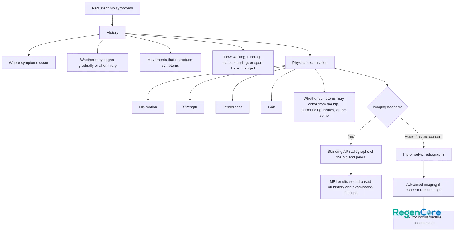
A clinician assessing persistent hip symptoms typically begins with the story rather than with an assumption about one structure. Relevant details include where symptoms occur, whether they began gradually or after an injury, which movements reproduce them, and how walking, running, stairs, standing, or sport have changed.
Imaging is selected for the clinical question
Imaging may be used when it is needed to clarify the suspected cause. For undifferentiated chronic hip pain, clinicians commonly begin imaging with standing anteroposterior radiographs of the hip and pelvis, then may consider MRI or ultrasound according to the history and examination findings. This is clinician-directed decision-making, not a sequence for self-ordering tests to prove a ligament diagnosis.
When there is acute pain and a possible fracture, hip or pelvic radiographs are generally appropriate initial studies. A normal X-ray does not always exclude an occult fracture. If clinical concern remains high—particularly after trauma or when a person who was previously walking normally cannot bear weight—advanced imaging may be needed, with MRI serving as the reference standard for occult hip fracture assessment.
Imaging can help answer a clinical question when selected in context. Persistent hip pain deserves a careful evaluation because anatomy alone, pain location alone, and a single test alone may point in the wrong direction.
When Hip Pain Needs Prompt or Urgent Assessment
Not every sore hip requires urgent care, but persistent or changing symptoms should not be dismissed as a presumed tendon or ligament issue.
Arrange medical assessment for persistent or activity-limiting symptoms
A medical assessment is appropriate when hip pain is persistent, worsening, recurring with a specific activity, or interfering with walking, standing, training, or daily life. It is also appropriate when symptoms include loss of motion, stiffness, swelling, redness, weakness, instability, nighttime pain, or popping, clicking, or catching. These signs do not identify the cause, but they provide a reason to look beyond self-management.
Seek urgent assessment for warning signs
Seek urgent assessment for inability to bear weight, severe uncontrolled pain, suspected fracture, or possible joint infection. Following trauma, a sudden inability to walk normally or bear weight needs timely evaluation, even if an initial X-ray is normal.
Acute hip pain with infection risk factors also needs urgent assessment. In people with risks such as poorly controlled diabetes, injection-drug use, or immunosuppression, the absence of a measured fever does not rule out concern for a joint infection.
These are general safety boundaries, not individualized triage. When symptoms are severe, rapidly worsening, or paired with the warning signs above, seek urgent medical care.
Arrive Ready for an Informed Hip Evaluation
The iliofemoral ligament is an important stabilizer at the front of the hip capsule, particularly as the hip moves into extension. Still, that anatomy is only one part of understanding hip symptoms. A productive evaluation focuses on the whole pattern rather than trying to decide in advance whether the issue is a ligament, tendon, joint, spine, or referred-pain problem.
Before your visit, make a brief record of:
- Exact symptom location: front/groin, outer hip, back/buttock, or more than one area.
- Onset: gradual, sudden, or tied to a fall, twist, collision, or other injury.
- Provoking movements: walking, running, stairs, prolonged standing, getting up from a chair, hip extension, rotation, or a sport-specific movement.
- Activity limits: what you have stopped, reduced, or modified because of symptoms.
- Motion and stability changes: stiffness, reduced range of motion, weakness, giving way, or a feeling of instability.
- Mechanical symptoms: clicking, popping, catching, or locking sensations.
- Prior history: previous hip, back, pelvic, or lower-extremity injuries and any prior imaging or treatment.
Bring these details to a physician evaluation, especially if symptoms are persistent or affecting activity. They help turn a vague concern about “hip pain” into a focused discussion of the likely sources and appropriate next steps for your situation.
Iliofemoral Ligament FAQ
What is another name for the iliofemoral ligament?
It is also called the Y ligament of Bigelow. The name reflects its usual Y-shaped branching form at the front of the hip capsule.
Where does the iliofemoral ligament attach?
It runs from the anterior inferior iliac spine of the pelvis to the intertrochanteric line of the femur. In some descriptions, its branches spread into an inverted Y as they attach along that region.
What does the iliofemoral ligament do in the hip?
Its main role is to reinforce the front of the hip capsule and limit excessive extension. It also helps support stability during extension and external rotation.
Can hip pain symptoms confirm an iliofemoral ligament problem?
No. Front, side, or back hip pain can come from several different structures, including the joint, tendons, spine, or referred sources. Symptoms can suggest a pattern, but they do not identify this ligament on their own.
What should I bring up during a physician visit for persistent hip pain?
Be ready to describe the exact pain location, when it started, whether there was an injury, which movements make it worse, any stiffness or loss of motion, clicking or catching, instability, and any prior hip or back injuries.
What imaging is usually considered for ongoing hip pain?
A clinician commonly starts with hip and pelvic radiographs when imaging is needed, then may consider MRI or ultrasound based on the history and examination. If fracture is suspected after trauma, a normal X-ray may not be enough to rule it out.